Muscles of the head and neck
The muscles of the head are small and there are two groups: the chewing muscles and the mimic muscles.
Chewing muscles: they enable chewing food when the mandible nears the superior maxillary (to close the mouth). They are short and wide and the most important are the temporal (it elevates the inferior maxillary) and the masseter (it elevates the inferior maxillary and grinds the teeth).
– Mimic muscles: they are located specifically in the face and enable gesturing. They connect with the skin and are flat and thin. The most important are: the frontalis (wrinkles the forehead and elevates the eyebrows), orbicularis of the eyelids (open and close the eyelids), the dilatator of the wings of the nose (moves the wings of the nose), orbicularis of the lips (move the lips), the risorius (retracts the corners of the mouth).
The muscles of the neck are numerous, thick and resistant, allowing the mobility of the head and maintaining an upright posture.
Among them we find the following groups:
– Lateral muscles of the neck: they are on both sides of he neck, in a symmetrical shape. One of the most important is the sternocleidomastoid, that flexes the neck and turns the head.
– Hyoid muscles: they are called this way for being in the hyoid bone region (throat), and their role is to lower and raise said bone, lower the pharynx and the mandible. Among them are the mylohyoid, the stylohyoid and the sternohyoid.
– Muscles of the nape: they are found in the back of the neck. Here we have the splenius, that extends the head and neck.
Muscles of the thorax and abdomen
The main muscles of the thorax are the pectoralis (major and minor), the intercostal (internal and external) and the serratus anterior.
– Pectoralis major and minor muscles: the first is located between the midline of the thorax and the axilla, and their function is to bring the arm closer to the trunk. The pectoralis minor is deeper and goes from the first ribs until the scapula bone and its function is to enable the traction of this bone.
– External and innermost intercostal muscles: these join the ribs to each other and to the sternum. In the case of the external intercostals, they elevate the ribs and help inspiration (inhaling air) and innermost intercostals lower the ribs and ease exhalation (expelling air).
– Serratus anterior muscles: they are found in the lateral walls of the thorax and join the first nine ribs to the scapula, making the shoulder move.
It must be pointed out that between the thoracic and abdominal cavity, inside the body, there is a muscle known as the diaphragm. It contracts when we inhale air, pushing up the ribs and increasing the volume of the rib cage.
Beneath the rib cage are the abdominal muscles, which, besides enabling the movements of the trunk, keep the inner organs (viscera) in their place and protect and compress them whenever it is necessary. In the abdomen we find several important muscles: the oblique (external and internal), the rectus and the transverse.
– Internal and external oblique muscles: they are born in the ribs and end in the bones of the hip. The external oblique muscle allows the tension of the abdomen and the inclination of the spine. The internal oblique tenses the abdominal wall and performs the flexion and rotation of the spine.
– Rectus muscle of the abdomen: it covers the area of the stomach and is located from the ribs to the pubis. It is used to flex the trunk forward and favors exhalation.
– Transverse muscle of the abdomen: it is the deepest and takes up the internal face of the abdomen. Its contraction unleashes an increase of the pressure in the abdominal cavity, contributing to exhalation, digestion, miction, etc.
Muscles of the back
The muscles of the back (or dorsal region of the trunk) are the ones that run the length of the spine and are numerous (superficial and deep). These are the most powerful muscles of the body because they enable us to stand, stay upright and lift and push objects.
Among the most important are: the trapezius, the posterior serratus and the latissimus dorsi.
– Trapezius: very large superficial muscle that takes up practically the center of the spine on both sides (from the cranium to the last dorsal vertebra). It enables us to elevate the shoulder and lift and rotate the scapula bone.
– Posterior (or dorsal) serratus muscle: they go from the central line of the body to the ribs, making them go up and down.
– Latissimus dorsi muscle: it is wide and long and is located in the inferior area of the back; it allows us to bring our arms backwards or inwards.
The rhomboid muscles (major and minor), the round muscles (major and minor), the infraspinatus and the supraspinatus are located in the scapular area and relate to abduction and rotation action of the scapula and the rotation of the arms. For this reason they are considered both muscles of the back as of the arms.
Other muscles that we will not get into in depth, but are considered of the back are the longissimus dorsi, the iliocostal, the posterior deltoids and the quadratus lumborum.
Muscles of the upper limbs
The main muscles of the upper limbs are grouped in the following areas: the shoulder, the arm, the forearm and the hand.
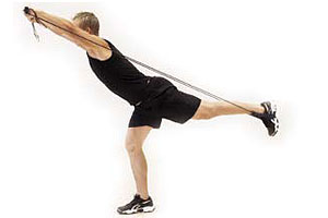
– Muscles of the shoulder: see box
– Muscles of the arm: they are two antagonistic muscles (they carry out contrary functions in order to enable a movement). One of them is the biceps brachialis (in the anterior part of the limb), which allows us to elevate the arm forwards and bend the elbow, elevating the forearm. The other is the triceps brachialis (in the posterior part), that extends the forearm and lowers the entire arm.
– Muscles of the forearm: they are in charge of moving the hand, flexing and extending the fingers. The round pronator flexes the forearm and turns the hand inwards. The supinators (long and short) enable the forearm to bend and the hand to twist outwards.
The palmaris longus (in the anterior part) flexes the forearm and hand. Finally, there are the deep and superficial flexors, which enable the flexing of the wrist and fingers.
– Muscles of the hand: they are short and small muscles that are in charge of flexing and stretching the fingers. The most important is the one that enables the opposition of the thumb; in other words, the «tweezer» action of the hand.
Muscles of the lower limbs.
These present a great development for they allow us to maintain the upright position, perform locomotion (walking and running) on the feet and sustaining the weight of the body. They are divided into the following groups; the hip and pelvis, the thigh, the leg and the foot.
– Muscles of the hip and pelvis: there are the ones of the posterior face, which include three muscles (gluteus maximus, gluteus median and gluteus minimus) that make up the buttocks, These have as their main function to extend the thigh and bring it closer to the midline of the body. In this area the psoasiliac muscles also stand out.
In the anterior area of the pelvis we find, among others, the long abductor that is born near the pubis and ends in the femur bone, and makes the leg go down and rotate, and the obturators inside the pelvis and serve to turn the leg outward.
Muscles of the thigh: the most prominent muscles of this area are quadriceps femoris and the biceps femoris (crural). The first is an extensor of the leg, and the second is an antagonist to the first (flexes the leg). In the thigh we also find the abductors, which are a group of fan-shaped muscles that ease the flexion and extension of the thigh.
Another prominent muscle is the sartorius (it is born in the bones of the hip and crosses the thigh obliquely) and are the ones that enable the crossing of the legs.
– Muscles of the leg: in its anterior part (above the tibia bone and until the foot) we find the anterior tibial muscle, whose action consists of turning the foot inwards. In the posterior part, the gastrocnemius (internal and external) are found superficially. They are born in the inferior area of the femur bone and are inserted below the achilles tendon.
The gastrocnemius act in collaboration with the soleus muscle to elevate and extend the heel.
– Muscles of the foot: see box.