Bodily functions demand that muscles carry out different tasks. For this reason there are three different kinds: voluntary muscles (or striated or skeletal), involuntary (or smooth) muscles and cardiac (or myocardial) muscle.
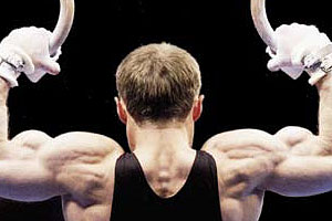
– Voluntary muscles: they hold the skeleton together (that is why they are also known as skeletal) with the help of the tendons. They are the ones that shape the body and help it with everyday movement.
Voluntary muscles are also called striated because they are made up of fibers (cells), that have horizontal strips (strias) that can be seen with a microscope. Their fibers are also characterized for being narrow, long and grouping in the thousands or hundreds. Each one measures from 1 mm to 4 cm in length and a few thousandths of a millimeter wide.
They have all the normal components of any other cell of the body, but they present two differences: one of them is that their number of mitochondria (where cellular respiration takes place) is greater because they must supply the great amount of energy the muscle needs, and the other difference lies in the existence of myofibrils, also in a number much greater than normal (several hundred).
The voluntary muscles (they are called like this because we can control their movements) are the majority because they are 600 of the 650 muscles present in the body. They are the ones that enable us to perform the locomotive function, in which the osseous system is the passive (support) component and the muscles are the active one, due to the fact that they contract, generating movement.
Since voluntary muscles are joined to the bone, they are mainly located in the legs, arms, abdomen and chest. They are also characterized for being able to contract quickly and strongly, that is why they wear out easily and have to rest between efforts.
– Involuntary muscles: they are made up of spindle-shaped (narrow and elongated), smooth looking cells (hence their name). This last bit because they lack transverse strias, although they show weak longitudinal strias.
They are characterized for their involuntary action (reason for which they are named so), which is activated by the nervous system and hormones. In the contraction itself, these muscles work in a way similar to the skeletal ones, but they take longer to contract. In addition, the involuntary muscles can remain contracted longer, because they do not wear out easily.
Smooth muscles are located in the internal organs (from here they also get the name «visceral») and in the large blood vessels. Therefore, the walls of the stomach and intestines are examples of these muscles, because they enable the decomposition of food and they move it through the digestive system.
– Cardiac muscle: it is located in the walls of the heart, enabling the rhythmic and powerful contractions that force blood outside this organ to take place. This muscle presents special characteristics because it could be said that its structure is striated, but its contraction is involuntary. However, some specifications must be made in both aspects.
In the case of the appearance of the fibers that make it up, although imperfect longitudinal and transverse strias are present in their cells, they differ from skeletal or striated muscle, especially in the central position of the cellular (or fiber) nucleus and in the ramification of the fibers.
In addition, the muscle fibers of the heart have a greater number of mitochondria, for the heart must not stop working. Regarding contractions, there is a difference in the traffic of nerve signals (between the muscle and the nervous system) because these have to be more continuous than frequent (if they were frequent the heart could wear out and die). Also, this muscle, unlike the striated and smooth, requires one to five seconds to contract again.
Other muscular classifications
In addition to the differences in structure among muscles, distinctions are also made according to their dimensions, shapes and location, among other aspects. Next, we will see other ways muscles are classified. According to their dimensions and shapes, muscles are classified into:
– Long: they are stretched and narrow and have great power. At the same time, they can be fusiform or flat, if their transverse diameter is greater in their middle part than at the ends. This way, the biceps is a long, fusiform muscle, while the rectus abdominis is long and flat.
– Short: they are those that, regardless of their shape, have very little length. For example, the ones of the head and face.
Orbicular: Their shape can be more or less circular and they are the ones that surround an important structure, like the ones in the mouth and eyelids.
– Wide: They are those whose diameters are, approximately, the same length and are generally flat and thin. An example is the lattisimus dorsi of the back. The muscles of the limbs can perform different movements, among these, those of flexion or extension, of rotation (pronation and supination), of approximation (adduction) or estrangement (abduction). This determines another typology of muscles, the categories of which are:
Flexors: they enable the flexion of the limbs (for example, bending the leg over the thigh or the arm over the forearm).
Extensors: they enable the extension of the limbs (example, stretching the leg over the thigh).
Pronators: they are the ones that make the extremities turn (for example, the hands) inwards.
Supinators: they are the ones that enable the outward inclination of the limbs.
Abductors: they are the ones in charge of moving the limbs away from the central axis of the body. An example is lifting an arm to the sides.
Abductors: These draw the limbs towards the central axis of the body. Example: putting the elbow at navel level.
As we have seen, most of the muscles are provided with tendons, through which they are usually inserted onto the bones. According to the type of insertion, meaning, if they do it through more than one end or head, they are divided into biceps (two heads), triceps (three heads) and quadriceps (four heads).
Depending on whether they are made up by more than one body or muscular belly, they are divided into digastric and polygastric (two or more bodies, respectively). If they take their terminal insertion by more than one end or tail, the muscles will be bicaudal, tricaudal or polycaudal, according to whether they do it by two, three or more ends.


 Día del Carabinero