The reproductive journey begins at the vagina, place where semen is deposited. From there, the sperms of semen enter the uterus through the cervix and then continue on to the fallopian tubes, precise spot of fertilization. The tale of spermatozoa eases their movement, but different contractions of the walls of the vagina and uterus also help. Only approximately two hundred sperms reach the reproductive encounter. They surround the ovule and start releasing a proteic enzyme called hyaloronidase, which modifies the surface of the female gamete, breaking up the layers that make it up. A single one is able to enter completely; when this happens, it activates substances that prevent entry to the other male gametes. A group of reactions and divisions come up next.The winning spermatozoon opens up a path through the different layers that cover the ovule, loses its tail and advances towards the encounter with the nucleus. Fertilization takes place when the nuclei fuse, the spermatozoon’s as well as the ovule’s. This way, 46 chromosome are assembled (each sexual cell contributes with 23), which is the new being’s share of chromosomes. Although half of the genetic information comes from each parent, the bond generates the creation of a unique and unrepeatable individual, which is now dubbed zygote.
Division and implantation
After fertilization, the zygote slowly descends along the fallopian tube in order to reach its final destination: the uterus. Since the new cell lacks movement of its own, it is helped by the cilia, or micro villi that lines the inside of the tubal mucosa, which move from side to side like arms, pushing it towards the uterine wall. During this journey, which lasts nearly eight or nine days, the zygote goes through several changes. Although it already contains all of the genetic material necessary to develop, it continues dividing and modifying. This way, approximately 36 hours after the encounter the new cell spits into two and performs this process through mitosis, forming two new identical cells, each one of them have an exact copy of the original cell’s genetic information. From there on out, a successive cellular division ensues, continuously doubling the new individual’s amount of cells. After 72 hours, the zygote will already be made up of 16 cells, acquiring a shape similar to that of a blackberry, and therefore, receives the name of morula. It ends its journey through the fallopian tube and finally reaches the uterus.However, the process of cellular division does not end there. When the morula becomes a more solid mass, now made up of 64 cells, it becomes a blastocyst or blastula. The blastocyst is a cellular mass (embryoblast) covered by an external layer (trophoblast), also made up of a liquid-filled cavity (blastocele). It is considered the stage prior to the development of the embryo, because it will definitely be the one implanted in the uterine walls. Approximately nine days after fertilization, the blastocyst definitely settles, taking advantage of the relaxation of the endometrium and the secretion of some enzymes from the trophoblast, which ease its implantation. The trophoblast will serve as the base for the growth of an important exchange substance (placenta), while the cells that make up the embryoblast will turn into the embryo.
Differentiation and formation
Once the blastocyst is implanted in the uterine wall, another important stage begins. But, unlike the previous ones, this period will be fundamental in the formation and location of the structures that will make up the anatomy of the new individual. The specific cells of each of the body’s systems begin to differentiate and take their location. For example, those that will make up the skeleton head outwards, while the cells that will form the internal organs take their place inside. Ten days after fertilization, the embryo’s appearance is still similar to a spherical cellular mass, but already has an important protective layer: the chorion. Up to the 15th day, three important germinative layers are developed, which will be fundamental for the growth of tissues, organs and structures that will form the new individual. They are the mesoderm, endoderm and ectoderm. The endoderm (also called entoderm) is the innermost of the three layers of the embryo, located in its abdomen. Structures as important as the digestive tract (from the pharynx to the rectum) are generated from it, as well as the respiratory organs and some glands like the liver, pancreas and thyroid gland. The middle layer is called mesoderm.Thanks to it, the dermis is spawned, along with the conjunctive tissue, vascular and urogenital systems and skeletal ad smooth muscle.
Finally, the ectoderm is the outermost of the three layers and develops on the dorsal side (back) of the embryo, originating the nervous system, epidermis; as well as hair and the crystalline of the eye. As of the third week of gestation, one of the most important systems when it comes to adaptation and survival is developed, when the embryo reaches 1.5 centimeters in length. It is the nervous system, which develops from the neuroblasts of the neural tube.Neuroblasts are primitive nerve cells that contain information that is fundamental for the development of he brain and the rest of the nervous system; they are found in the neural tube, which is derived from the ectoderm. The encephalon and cranial nerves are formed from its superior part, while the inferior portion forms the spinal cord.
Microscopic growth
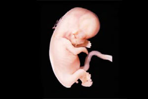
During the first months of life, the embryo clearly does not have a human appearance. From being a small cellular sphere, as of the first two weeks of gestation it evolves until it acquires, at the end of the first month, a shape similar to a larva. It is possible to make out the outline of a head, a small curved tail (which will disappear in time) and, in its middle portion, a budding heart, which functions as propulsion pump as of the 22nd day. Its hardest portion is located on the external side of the embryo, a small outline of what the spine will be. In the second month, the embryo develops anatomical structures from where the ears, eyes, kidneys, arms and digestive and circulatory systems will grow; the umbilical cord reaches 12 cm in length, being the only structure the embryo has to communicate with the placenta. At the end of this month, the embryo can measure up to four centimeters and weigh close to four grams. Although its size is small, it acquires a human form little by little. It even already has most of its organs under development, each one positioning itself correctly.


 Muere Evita